


1 September 2026
No Comments
iHuman assignment
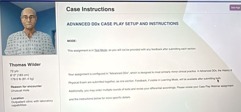
Thomas Wilder
72 y/o
6′ 0″ (183 cm)
179.0 lb (81.4 kg)
Reason for encounter
Unusual mole
iHuman Thomas Wilder unusual mole History Questions
- How can I help you today?
- Do you have any other symptoms or concerns we should discuss?
- Do you have a mole that has changed?
- When did you notice the change in your mole?
- Have you had any skin sores or change in skin color?
- Do you have any skin itching or burning?
- Can you tell me about any current or past medical problems you have had?
- Have you had any recent blood or lab tests? What were the results?
- Any previous medical, surgical, or dental procedures?
- Are you taking any prescription medications?
- Are you taking any over-the-counter or herbal medications?
- Do you have……..
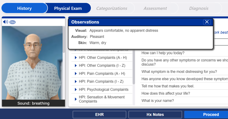
iHuman Thomas Wilder unusual mole answers
Physical Exams Required
- Weight
- Height
- cognitive status
- SpO2
- temperature
- blood pressure
- pulse
- respiration
- temperature
- auscultate heart
- auscultate lungs
- assess …….
Unpacking the iHuman Case Study: 72-Year-Old Thomas Wilder and the Evaluation of an Unusual Mole
Navigating clinical simulation modules on iHuman requires a structured, evidence-based approach to patient assessment. When managing complex dermatology patient encounters—such as the clinical presentation of Thomas Wilder, a 72 year old male presenting with an unusual mole—nursing students and advanced practice clinicians must synthesize subjective history, detailed physical findings, and risk factors to formulate an accurate clinical picture.
This case study breakdown provides an strategic overview of the clinical assessment process, key evaluation metrics, and general diagnostic pathways involved when evaluating suspicious skin lesions in older adults.
Patient Overview: Meet Thomas Wilder
In this iHuman scenario, students are introduced to Thomas Wilder, a 72 year old retired farmer who presents with a chief complaint of a newly noticed, evolving skin lesion on his right shoulder. The initial presentation highlights several critical historical factors:
Demographics: 72-year-old male with fair skin (Fitzpatrick Type II).
Occupational History: Decades of agricultural outdoor work leading to significant cumulative solar UV exposure.
Primary Concern: A dark brown, scaly lesion on the right shoulder that has changed in texture and border over the last several months and causes localized discomfort when rubbing against clothing.
Recognizing how occupational hazards like chronic sun exposure intersect with aging skin is essential for guiding your clinical reasoning throughout this module.
Conducting the Subjective History: Essential History Questions
Obtaining a targeted history of present illness (HPI) is the cornerstone of any dermatological evaluation. When completing history questions in virtual patient simulations like iHuman, clinicians must methodically explore the characteristics of the chief complaint.
Key areas of inquiry when evaluating an unusual mole include:
Symptom Onset and Chronology: Determining exactly when the lesion appeared and how rapidly it has evolved over time.
Symptom Progression: Asking about alterations in size, shape, surface texture, or color intensity.
Associated Symptoms: Identifying local mechanical irritation, pruritus, tenderness, bleeding, or exudate.
Risk Factor Assessment: Gathering details on personal and family histories of skin malignancies, past severe sunburns, and total lifetime UV exposure.
Systemic Review: Ruling out B-symptoms such as unexplained weight loss, night sweats, or localized lymphadenopathy.
Mastering the structure of these patient interview questions ensures that no critical subjective clues are missed during your simulation grading.
The Physical Examination: Objective Evaluation Strategies
A thorough physical examination in dermatology relies on systematic inspection, palpation, and specialized clinical diagnostic tools. In the assessment of Thomas Wilder, objective examination techniques help distinguish benign epidermal growths from potentially malignant cutaneous lesions.
Key Components of the Dermatological Exam:
Inspection & Visual Assessment: Evaluating the symmetry, margin definition, pigmentation uniformity, and overall diameter of the lesion.
Palpation: Checking for elevated surfaces, hyperkeratotic scale, firmness, or underlying induration.
Dermoscopic Evaluation: Utilizing dermoscopy to identify micro-structural patterns such as keratin pseudocysts, comedo-like openings, pigment networks, or atypical vascular arrangements.
Full-Body Skin Survey: Inspecting surrounding sun-exposed and non-exposed anatomical sites for dysplastic nevi, actinic damage, or secondary lesions.
Regional Lymph Node Assessment: Palpating regional nodal basins (cervical, axillary, and supraclavicular) to assess for lymphadenopathy.
Formulating a Differential Diagnosis
Developing a robust differential diagnosis requires cross-referencing subjective risk factors with objective skin patterns. For a 72 year old patient with chronic UV exposure presenting with an unusual mole, several key diagnostic possibilities must be considered:
Seborrheic Keratosis: Common benign epidermal tumors characterized by a “stuck-on” appearance, waxy texture, and dermatoscopic pseudocysts.
Actinic Keratosis / Squamous Cell Carcinoma in situ: Precancerous or early malignant lesions presenting as rough, scaly, hyperkeratotic plaques on sun-damaged skin.
Malignant Melanoma: A high-risk cutaneous malignancy evaluated using the standard ABCDE criteria (Asymmetry, Border irregularity, Color variation, Diameter >6 mm, Evolving).
Basal Cell Carcinoma: Translucent, pearly, or pigmented nodular lesions often displaying telangiectasias or central ulceration.
Evaluating these conditions side-by-side allows clinicians to justify whether advanced diagnostic testing (such as a shave or punch biopsy) is necessary.
Developing a Comprehensive Management Plan
Once the primary working diagnosis is established, a patient-centered management plan must be constructed. In clinical practice and iHuman grading rubrics, management plans encompass several distinct domains:
Diagnostic Testing & Biopsy Decisions: Determining whether observation, dermoscopic monitoring, or immediate tissue biopsy is clinically indicated.
Therapeutic & Symptomatic Interventions: Offering topical barrier creams, short-term low-potency anti-inflammatory treatments for friction relief, or cryotherapeutic options if requested for cosmetic or functional comfort.
Patient Education & Prevention: Advising on broad-spectrum sunscreen application (SPF 30+), protective clothing, self-skin examinations, and regular primary care surveillance.
Specialty Consultations: Identifying appropriate thresholds for specialty dermatology referrals.
Follow-Up & Red Flag Counseling: Establishing follow-up timelines and educating the patient on warning signs (such as rapid growth, color changes, or spontaneous bleeding) that require prompt medical re-evaluation.
Get Complete, Top-Graded Solutions for Your iHuman Cases!
Are you working through the Thomas Wilder case or other complex iHuman clinical scenarios? Achieving a perfect score on your history taking, physical exam selection, differential prioritization, and management plan documentation can be challenging.
Don’t leave your grades to chance! At iHuman Tutor, we provide comprehensive, step-by-step case guides, fully developed HPI rubrics, exact ROS checklists, complete physical exam findings, and evidence-based management plans tailored for nursing students and clinicians.
👉 Ready to excel in your coursework? Visit iHuman Tutor today to order expert-verified case solutions and boost your clinical confidence!